
The Site of Bone Marrow Acquisition Affects the Myeloid to Erythroid Ratio in Apparently Healthy
Myelodysplastic syndromes (MDS) are a group of myeloid neoplasms that are often difficult to diagnose due to their pathologic and clinical heterogeneity. The key features of MDS are peripheral blood cytopenias, ineffective hematopoiesis manifesting as morphologic dysplasia, and clonal genetic abnormalities.

Thread by 4theLoveofPath Instead of my normal GTScase, I wanted to look at the morphology of
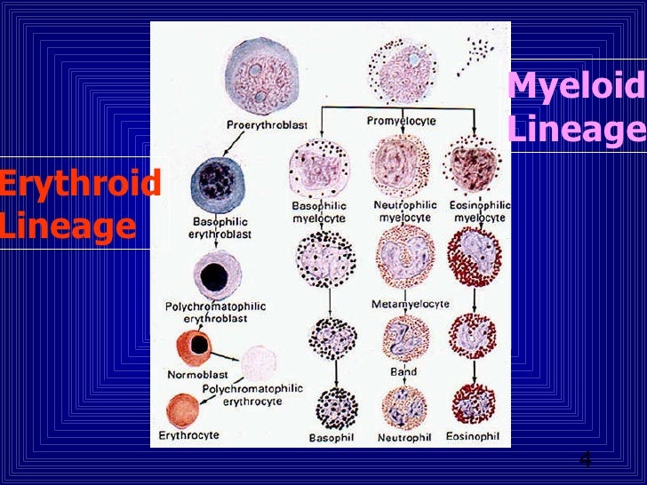
The early myeloid progenitors are localized in the paratrabecular areas close to the adventitia of the small arteries. Normally, the layer of immature granulocytes does not exceed 2-3 rows of maturing cells. With maturation, the cells migrate to the intertrabecular spaces.
Acute myeloid leukemia with expanded erythropoiesis Haematologica
A bone marrow biopsy showed increased cellularity (85%) , increased myeloid to erythroid ratio, myeloid left-shift, and a markedly increased number of megakaryocytes varying from atypically small.
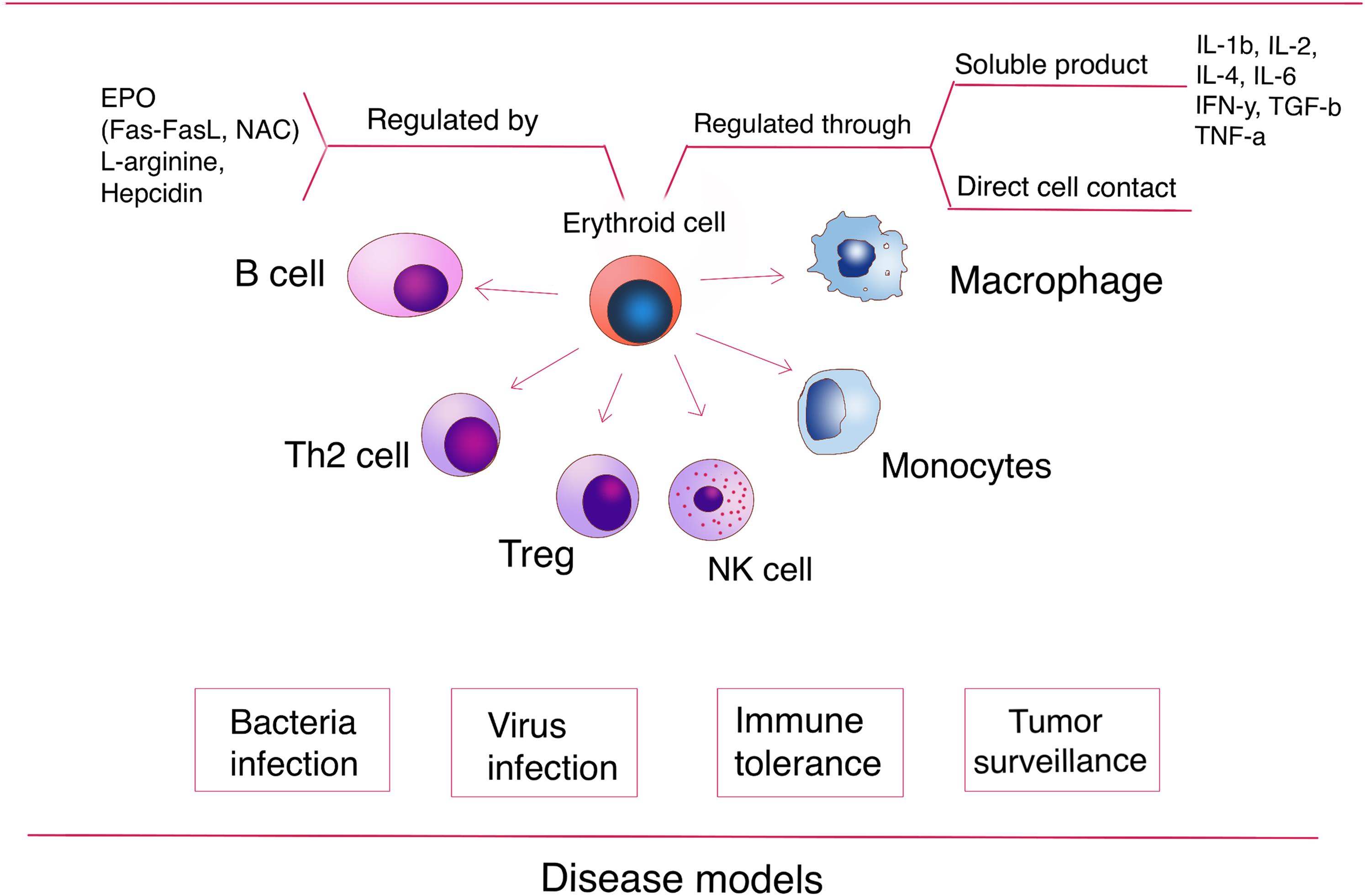
Erythroid Lineage Cells in the Liver Novel Immune Regulators and Beyond
Introduction The bone marrow is the largest primary lymphoid organ and is one location of antigen-independent lymphocyte development. It is also a secondary lymphoid organ because terminal antigen-induced lymphoid cell differentiation occurs within its microenvironment ( Tavassoli and Yoffey, 1983 ).
PPT Bone Marrow Evaluation PowerPoint Presentation, free download ID671342
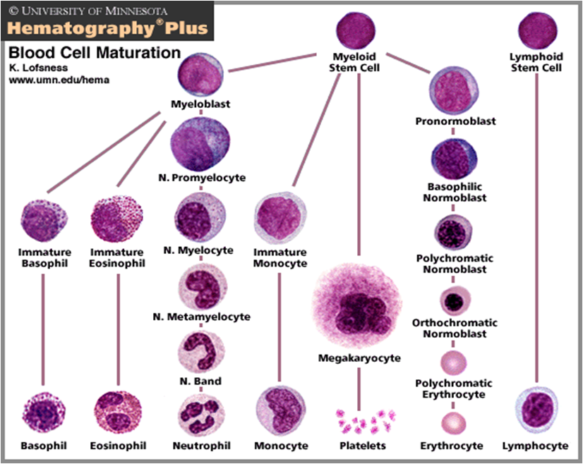
Hematopoietic components Bone marrow aspirate showing normal "trilineage hematopoiesis": myelomonocytic cells (an eosinophil myelocyte marked), erythroid cells (an orthochromatic erythroblast marked), and megakaryocytic cells

Advances in understanding erythropoiesis evolving perspectives Nandakumar 2016 British
Normal M:E Ratio The normal M:E ratio in adults varies from 1.2:1 to 5:1 myeloid cells to nucleated erythroid cells. An increased M:E ratio (6:1) may be seen in infection, chronic myelogenous leukemia or erythroid hypoplasia. A decreased M:E ratio (<1.2-1) may mean a decrease in granulocytes or an increase in erythroid cells.

Myeloid to erythroid ratio (ME ratio) presented as mean and... Download Scientific Diagram
The benefits to core biopsy evaluation is a more accurate representation of bone marrow cellularity and tissue architecture (such as fibrosis, focal neoplastic infiltrate, necrosis, etc.) than aspirate and cytology. However, cell morphology is more difficult to assess.

Myeloid to erythroid ratio eClinpath
Myeloid and erythroid cells: With myeloid and erythroid precursors, we do the following: 1) Assess for complete and balanced maturation, 2) Calculate a myeloid to erythroid ratio, 3) Evaluate morphologic features and 4) Look at cell proportions.

Fat , Myeloid , Erythroid cell ratio in bone marrow (*) FaME Bone Marrow Cells, Hematology
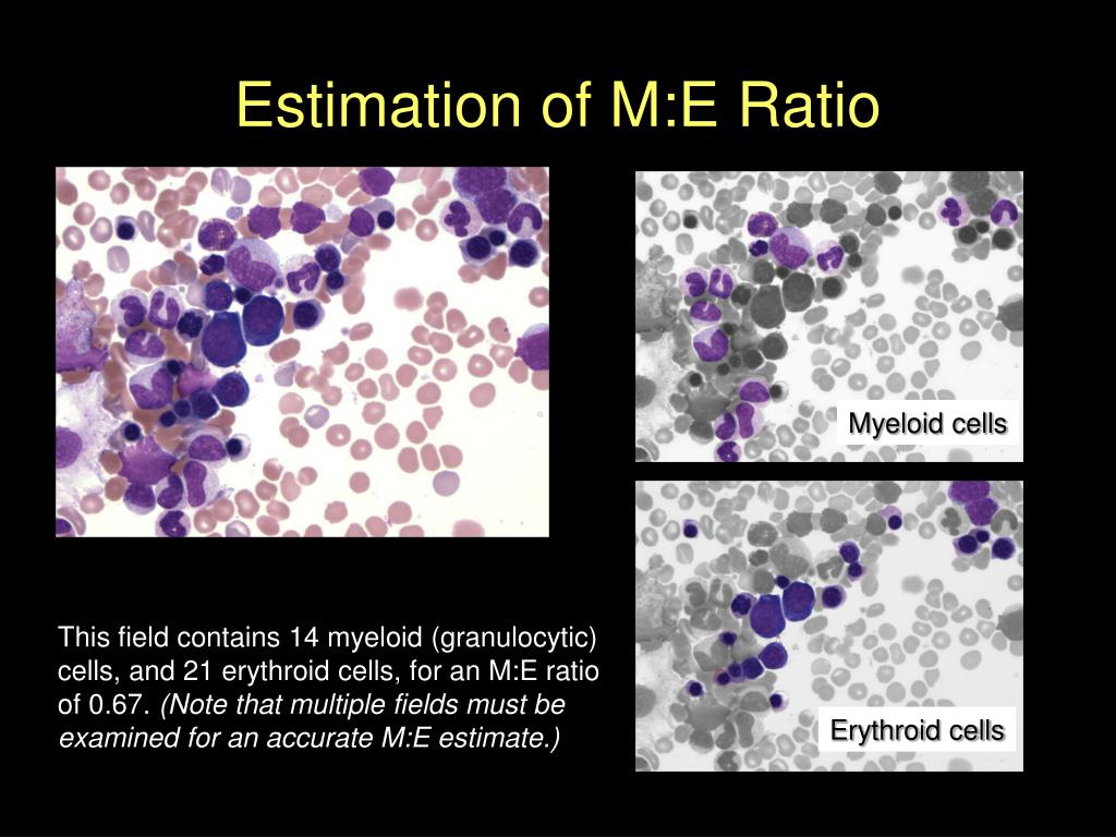
7 Estimation of myeloid:erythroid ratio comparing granulocytes and erythroid precursors.. Erythroid, myeloid, and/or megakaryocytic hypoplasia are terms applied to the situation in which there are fewer precursor cells than appropriate for the number of mature cells in peripheral blood. For example, the absence of erythroid hyperplasia in an.

Immunology What Cells Have a Myeloid Lineage and How Are they Identified?
Marrow aspirate specimen with a myeloid/erythroid ratio (M/E ratio) of 1:1-2, typical for a patient with a hemolytic anemia or one recovering from blood loss. View Full Size | | Download Slide (.ppt) + + FIGURE A6-6. Myeloid hyperplasia of the marrow. Marrow aspirate specimen showing a myeloid/erythroid ratio of ≥3:1, suggesting either a.

Megakaryocyte number and myeloid erythroid ratio in intact control... Download Table
What is being tested? Bone marrow is the soft and sponge-like tissue found inside the body's larger bones that produces blood cells. Bone marrow aspiration and biopsy are procedures used to collect and evaluate bone marrow cells and structure.

myeloid erythroid ratio Ratio, Image, Map
Myeloid cells: Normal or abnormal maturation Indicate if excess blasts Abnormal localization of immature precursors Erythroid cell: Normal or abnormal maturation Provide differential of myeloid and erythroid elements, based on counting 200 - 500 cells in aspirate smear Megakaryocytes: Normal or abnormal numbers and morphology Lymphocytes:

PPT Myeloproliferative Disorder PowerPoint Presentation ID3066493
Myeloid to erythroid ratio By Tracy Stokol / January 1, 2019 In a normal bone marrow, there is an approximately 1:1 (ranging from 0.7:1 to 2:1) ratio of myeloid (M) to erythroid (E) progenitors in marrow (Wright's stain, 50x objective)
Rbc
Myeloid cells make up the largest percentage of the normal marrow cellularity; erythroid cells are second most common. The ratio of myeloid to erythroid cells should be about 2:1 to 4:1. It's easier to see these cells on an aspirate smear, but you can get a pretty good idea on the marrow section too.
Dentistry and Medicine CHRONIC MYELOID LEUKAEMIA
Myeloid:erythroid ratio | definition of myeloid:erythroid ratio by Medical dictionary Myeloid:erythroid ratio | definition of myeloid:erythroid ratio by Medical dictionary https://medical-dictionary.thefreedictionary.com/myeloid%3aerythroid+ratio Dictionary, Encyclopedia and Thesaurus - The Free Dictionary 13,715,621,931 visits served

PPT Bone Marrow Evaluation PowerPoint Presentation ID671342
Interestingly, a peculiarly normal cellularity and myeloid-to-erythroid (M:E) ratio was reported in seven (50%) and 11 (84.6%) out of the 14 and 13 patients with reported data, respectively. Moreover, megakaryocytes were small in 10 patients (71.4%), pleomorphic in three patients (21.4%), and dysplastic in a single patient (7.1%), respectively.